|
 |
INTRODUCTION
|
|
In this copy of the newsletter, we share an interesting story about a Rove beetle that was collected by Charles Darwin in 1832; announce our $250 imaging service prize winner from ESA; share our technical innovations; and share our most notable images from the past few months. We hope you enjoy reading the newsletter and we wish you a Merry Christmas and a happy New Year!
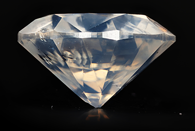
Fancy White Diamond, standing in clay. Milky White 1.08 ct 3x 1,16
|
|
NEWS
|
WINNER of $250 Imaging Service Voucher
Macroscopic Solutions offered attendees of the Entomology 2014 and the Entomological Collections Network 2014 meetings in Portland, OR a chance to win $250 worth of imaging services to one lucky newsletter subscriber. Macroscopic Solutions is happy to announce Shane J Burchfield of Bugs of America as the winner. Congratulations Shane! Macroscopic Solutions will be contacting you shortly.
|
|
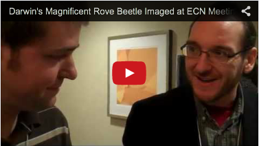
Charles Darwin’s Rove Beetle Imaged
Each year, a group called the Entomological Collections Network (ECN) meets to discuss topics about taxonomy, systematics, museum collections, interpreting data, and more. Plus, many of the attendees bring insects from all over the world, which they loan to other entomologists for research purposes.
This year a sample that was being described by Dr. Stylianos Chatzimanolis of the University of Tennessee was being returned to Dr. Max Barclay who is the Collections Manager of Coleoptera and Hymenoptera at the Natural History Museum in London.
Dr. Max Barclay generously brought the beetle to the Macroscopic Solutions exhibit where it was imaged with the Macropod. The sample required no preparation and the image was completed in less than 5 minutes.
|
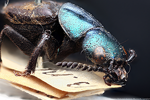
Darwins Rove Beetle
|
“This is a Staphylinid beetle, a rove beetle, collected by Charles Darwin on the voyages of the Beagle in modern-day Argentina,” Dr. Barclay said. “ “This one sat in the unidentified material for more than 150 years until Stelios Chatzimanolis came to London just a year ago, and he recognized it as a new species, and he asked whether he could borrow it so that he could take it away and describe it, and he named it after Darwin,” he continued. “The genus is Darwinilus … and he named the species after somebody called David Sedaris. It’s Darwinilus sedarisi.” |
Technical Innovations
The images Macroscopic Solutions typically show are considered macroscopic. Meaning the specimens are magnified by less than 10x; however, the resolution of the image will allow magnification up to 25x (Microscopic). Macroscopic Solutions has now far and away exceeded these limitations, as shown in the pictures below.
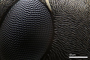
This is a typical Macropod image of the ant, Formica serviformica. It was shot with the Macropod at 5x magnification.
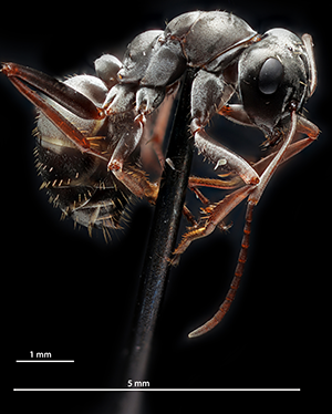
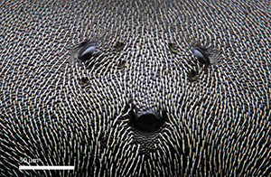
Now, 40x magnification is reachable, with the resolution allowing details to be seen at approximately 100x magnification. The main visual structure of an ant's compound eye (Formica serviformica) is comprised of many hundreds or thousands of tiny facets. Each facet is actually the corneal lens or an individual photoreceptor unit called an ommatidium. An ommatidium is divided into light gathering and light detecting components, which allow the insect to detect motion.
Three dorsal ocelli of the ant Formica serviformica. Dorsal ocelli are light-sensitive organs found on the dorsal or frontal surface of the head. Ants have ocelli and compound eyes, which means that they possess two anatomically separate and functionally different visual pathways. A dorsal ocellus consists of the cornea and a layer of photoreceptors. The refractive power of the lens is not typically sufficient to form an image on the photoreceptor layer, but the convergence ratio from the photoreceptor to the neurons is large.
|
| |
NOTABLE MACROPOD IMAGES
|
|
The Macropod’s images are sometimes considered a mixture of science and art. This is because a scientific instrument is used to achieve an artistic end that is significant for discovery, observation and education. The images appease the senses and document high quality data of subjects that are associated with time and position.
Macroscopic Solutions releases many images daily and the some of our most noteworthy are shown below.
|
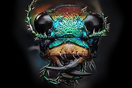
Tiger Beetle, Arizona, Cochise Co.
| 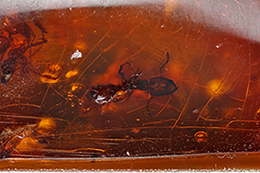
Ant in amber, Dominican Republic
|
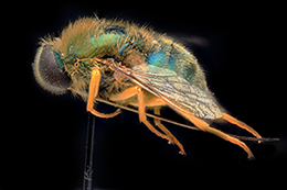
Eulonchus smaragdinus
|
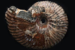 Pyritized ammonite Pyritized ammonite
|
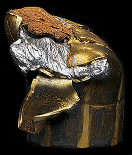
Remington golden saber 230 bullet with wood from tree
|

Citrine crystal
|
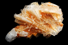
Strontianite crystals, Cedar Hill Quarry, Cedar Hill, PA
|
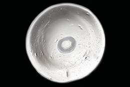
Contact Lens Negative
|
| |
EVENTS AND CONFERENCES
|
Since their formation, Macroscopic Solutions has exhibited their high-performance imaging equipment at a range of scientific conferences, led workshops on macro-imaging techniques, and delivered multiple presentations on scientific imaging. This provides researchers, museum collections managers, students, consultants, etc. to observe the techniques used by Macroscopic Solutions first-hand. Over the next few months, you can find Macroscopic Solutions at the following events and conferences. |
Macropod Presentation at the Smithsonian
On March 17, 2015, Macroscopic Solutions will be giving a presentation at the Smithsonian National Museum of Natural History on scientific imaging techniques that are made simple with their novel imaging system, the Macropod. The invitation was given by the Guild of Natural Science Illustrators (GNSI) and a range of scientific communities will be present, including the Entomological Society of Washington, DC, the Professional Photographer Society of Greater Washington, the Botanical Society of Washington, the Illustrators Club of Washington. This event is open to the public.
Schedule
5:30 – Meeting opens with socializing and snacks
6:00 – Announcements and GNSI business
6:15 – Macroscopic Solutions presentation |
Geological Society of America Northeastern Section
The GSA Northeastern Section is celebrating its 50th Anniversary in 2015, and to do so it is returning to The Omni-Mount Washington Resort in Bretton Woods, NH. The meeting is scheduled from March 23-25. Visit the GSA website for information on registration, symposia, the resort, and more. |
Northeast Natural History Conference
The 15th Northeast Natural History Conference (NENHC) will be held from April 18-20, 2015 at the Sheraton Springfield Hotel in Springfield, MA. This conference promises again to be the largest regional forum for researchers, natural resource managers, students, and naturalists to present current information on the varied aspects of applied field biology (freshwater, marine, and terrestrial) and natural history for the Northeastern United States and adjacent Canada. For more information about this event, visit the NENHC website. |
**FREE IMAGING SERVICES**
By showing this newsletter at these conferences, you will receive one free high-resolution image of your specimen on-site AND you will be entered for a chance to win $250 worth of Macroscopic Solutions imaging services. Click Here to read more about imaging services. |
| |
|
|
|